January 3, 2012

Proliferative enteropathy (PE), commonly referred to as ileitis, is an infectious, enteric disease characterized by thickening of the intestinal mucosa. Within these proliferating cells is Lawsonia intracellularis, an intracellular bacterium.
Two clinical forms, acute hemorrhagic diarrhea and chronic intestinal adenomatosis, have been historically well described; however, a subclinical form is currently the most commonly reported. Subclinical cases are mainly characterized by intermittent fecal shedding of the bacteria associated with poor growth rates and variations in market weights.
The presence of this bacterium in the hostile intestinal microenvironment and the lack of specific cell-free media or broth to support its isolation/growth in the laboratory have challenged many bacteriologists over the years. Therefore, isolation of the bacterium from fecal samples has not been a feasible approach to diagnosing the infection in living pigs.
Nevertheless, the University of Minnesota Veterinary Diagnostic Laboratory is one of the few laboratories worldwide which is able to isolate L. intracellularis in cell culture systems. Laboratory culture of L. intracellularis is important to develop alternative methods for diagnosis, to study susceptibility to different antimicrobials, and to investigate how ileitis causes disease.
Diagnosing Ileitis Infection
Postmortem diagnosis is based on lesions characterized by thickening of the intestinal mucosa mainly located near the ileal-cecal junction (Figure 1). In contrast with entero-colitis (caused by Salmonella typhimurium) or hemorrhagic colitis (dysentery, caused by Brachyspira sp.), inflammation is not the primary lesion of ileitis. This fact has complicated the diagnosis of ileitis infections in the field, especially in mild, chronic or subclinical cases. Additionally, Peyer’s Patches (mucosa-associated lymphoid tissue) present in the ileum gives an apparent thickened aspect to the mucosa and makes the macroscopic diagnosis even more subjective. Therefore, confirmation of the clinical diagnosis with laboratory techniques is warranted. Intestines of pigs affected by the acute form are dilated and thickened with a corrugated serosal surface. Blood clots may be found in the lumen of the ileum or colon (Figure 2).
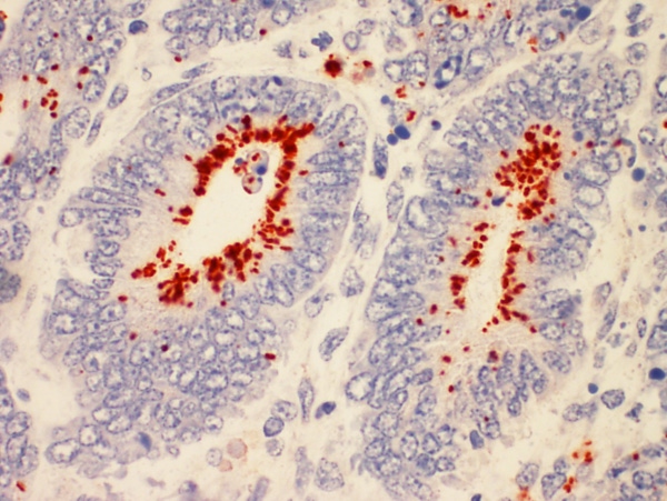
When intestinal tissues are collected from suspected ileitis cases, the diagnosis can be made microscopically and L. intracellularis infection can be confirmed through immunohistochemistry (Figure 3).
While this disease continues to be an important cause of diarrhea, the number of cases diagnosed with Lawsonia intracellularis infection at the Minnesota Veterinary Diagnostic Laboratory by immunohistochemistry has declined during the last few years (Figure 4). There are different factors contributing to this decline. On the one hand, vaccination and medication strategies have helped control this disease in some herds. On the other hand, the ability of veterinarians to diagnose this disease solely based on clinical signs and gross lesions has improved.
Finally, use of polymerase chain reaction (PCR) has increased, which can be done on feces from live animals. Herds can be monitored in the field by fecal PCR, which is the best option to identify animals actively infected. However, this approach is not sufficiently sensitive for the diagnosis of all infections, especially subclinical forms. Methods described for the serologic diagnosis have employed whole bacterial antigen incorporated into an immunoperoxidase monolayer assay (IPMA) and ELISA-based assays. These assays are more sensitive than PCR when used for cross-sectional or longitudinal monitoring of subclinically affected herds.
For definitive diagnosis, necropsy followed by specific identification of L. intracellularis in the lesions by immunohistochemistry, continues to be the gold standard.
Tracking Ileitis Infection
Little information is available on the source or spread of L. intracellularis within or between ileitis outbreaks. This fact compromises any efforts related to potential eradication of the disease in a production system. The University of Minnesota Veterinary Diagnostic Laboratory developed and is now applying a variable number tandem repeat technique for typing outbreaks. This technique identifies the DNA sequences of four regions in the bacterial genome and creates a specific Lawsonia profile for each outbreak. Furthermore, the technique is applicable to PCR-positive feces and tissue without the necessity of cultivating the bacteria, allowing sub-specific identification of field isolates.
We believe epidemiologic profiling is a promising tool for tracking outbreaks by identifying the Lawsonia strain from commonly sourced animals (replacement gilts) and their nursery and finishing sites. Control points may be identified in systems that supply breeding stock where downstream nursery and finishing sites are also experiencing variable levels of ileitis.
Click to view graphs.
Fabio Vannucci, DVM; Albert Rovira, DVM and Connie Gebhart
University of Minnesota Veterinary Diagnostic Laboratory
[email protected]
You May Also Like


