Subscribe to Our Newsletters
National Hog Farmer is the source for hog production, management and market news
Respiratory infections would include Actinobacillus pleuropneumonia and Actinobacillus suis. There are many individual pig events, such as electrocution, trauma, aneurysms, etc., that are not group issues
May 26, 2011

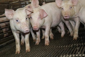
There are a variety of conditions that cause sudden death in finishing pigs. In general, sudden death in finishing pigs can be divided into enteric (gut) conditions, respiratory (lung) infections and individual pig events. Enteric conditions would include ileitis, hemorrhagic bowel syndrome (HBS), twisted gut and stomach ulcers. Respiratory infections would include Actinobacillus pleuropneumonia and Actinobacillus suis. There are many individual pig events, such as electrocution, trauma, aneurysms, etc., that are not group issues.
Acute ileitis can be seen throughout the finishing period, and should not be confused with chronic ileitis that causes diarrhea in all ages of finishing pigs. With acute ileitis, pigs may show no signs of diarrhea at all. If there is any diarrhea, it is a black, tarry feces and not the feed-colored diarrhea associated with chronic ileitis.
Acute ileitis is most commonly seen in heavyweight market animals, commonly after the first marketing from the group. Farms that see acute ileitis often see it repeated in subsequent groups if there is no intervention. Postmortem examination reveals pale pigs that appear gaunt with a thickening of the ileum. The ileum is usually filled with clotted blood.
Twisted gut and HBS are commonly diagnosed in growing and finishing pigs. Caretakers may walk through a group of pigs and return in 30 minutes to find a pig that has died, looking pale with a distended abdomen. In the case of pigs succumbing to a twisted gut, the intestinal tract turns on its base, much like twisting a towel to wring out water, which stops the blood supply to the intestinal tract.
On postmortem examination, both conditions yield pigs with intestines that are distended with unclotted blood and gas, but the twisted gut pigs have a knot or twist at the base of the intestines. With HBS, the intestinal tract is lying in its normal position. Some suggest that HBS pigs died from a “twisted gut” that untwisted after death.
Stomach ulcers can be very slow to develop or happen quite rapidly. In many chronic cases, clinical signs will consist of pigs losing weight. These pigs appear pale. With the sudden deaths due to ulcers, the ulcer erodes into a blood vessel and the pigs bleed out into the stomach. On postmortem examination, these are good-looking pigs with stomachs that are filled with a large blood clot, and die prior to passing significant amounts of blood into the intestinal tract.
Actinobacillus pleuropneumonia (APP) and Actinobacillus suis (AS) are bacterial infections of the lungs that cause sudden death in all ages of swine. Both organisms can cause numerous deaths at a single visit. With APP, it is common to see pigs that have recently died with blood coming from the nose. On postmortem examination, at least one lung will have a distinct hemorrhagic lesion. AS causes a more dispersed lung lesion, and the organism is found in many organs.
Case Study No. 1
We were called to a 4,800-head, wean-to-finish site consisting of two, 2,400-head barns with a history of multiple sudden deaths each of the prior three days. Ten pigs that appeared normal had died over the three days.
Postmortem examination revealed all three pigs had stomach ulcers containing large blood clots. Although the group appeared healthy, as we walked through the barn, this group had gone through an influenza infection the week before. The off-feed periods from the influenza are believed to have triggered the surge of ulcers. In the next couple of days, the mortality ceased.
Case Study No. 2
We were called to a 2,400-head, wean-to-finish site with a history of sudden death in late finishing. The pigs had been on feed for approximately 21 weeks. Two pigs had recently died in one pen. The pigs appeared pale and empty. Postmortem examination revealed pigs with empty stomachs and small intestines filled with clotted blood. The colons were filled with black, tarry feces. Other pigs in this pen had black feces over their rectal areas.
A diagnosis of acute ileitis was made and treatment was started. The group was started on tylosin in the water as per label directions and 100g/ton tylosin in the feed. The pigs with clinical signs recovered and the groups showed no further signs of ileitis.
In the case of sudden deaths, the pigs look normal after they die, so postmortem examinations are necessary to achieve a proper diagnosis. In the absence of clinical signs prior to death, working with your veterinarian to put in place prevention protocols is the key to a successful program.
You May Also Like
Enter a zip code to see the weather conditions for a different location.